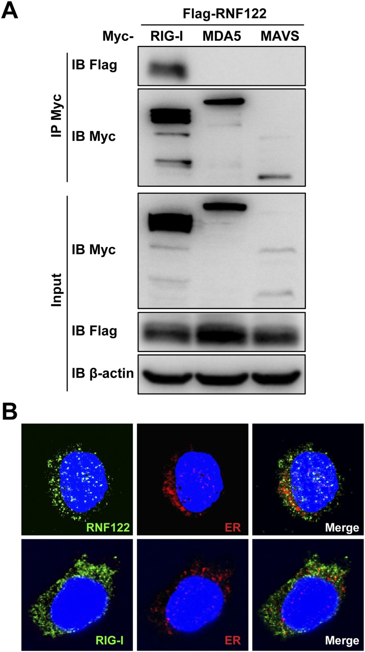
Fig. S1.
Interaction of RNF122 with RIG-I, MDA5, or MAVS. (A) The indicated plasmids were transfected into HEK293T cells. Protein–protein interactions were detected by immunoprecipitation using the anti-Myc antibody, followed by Western blot analysis using the indicated antibodies. (B) Peritoneal macrophages were infected with VSV for 8 h. Cells were then immunostained with anti-RNF122 (green) and the ER marker anticalreticulin (red; Upper), or anti–RIG-I (green) and the ER marker anticalreticulin (red; Lower) and analyzed by fluorescence microscopy. (Magnification, 400×.)

