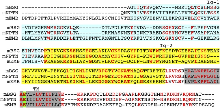
Fig. S4.
Alignment of the amino acid sequences of mouse basigin (mBSG), neuroplastin (mNPTN), and embigin (mEMB). The amino acid sequences of mBSG, mNPTN, and mEMB were aligned to obtain the maximal homology by introducing gaps (–). The amino acid residues that are identical among the three members are in bold red, and the residues that are conserved only between mBSG and mNPTN are in light red. The first (Ig-1) and second (Ig-2) Ig-like domain 1 are highlighted in light blue and yellow, respectively. The putative transmembrane regions are shadowed in gray, and the atypical glutamic acid residues in the transmembrane region are highlighted in green.

