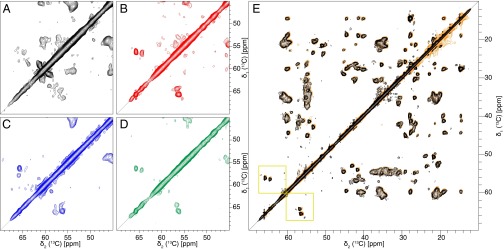
Fig. 1.
Screening of conditions toward a sample with a single polymorph and reproducibility of the sample preparation. The 2D [13C,13C] DARR spectra (20-ms mixing time, 13 kHz MAS, 14 T B0) of Aβ(1–42) fibrils grown at different conditions. A–D show the serine region used to determine the amount of polymorphs in the sample. Aβ(1–42) contains two serines (residues 8 and 26), but under condition 0 (A), at least six serines can be counted. Furthermore, under this condition, the lines are very broad, and no clear defined peaks are observable. Under condition 3 (C) the lines are narrower, but still about 6 serine peaks are visible. For conditions 2 (B) and 4 (D) a single set of resonances is observed, which indicates the presence of only one morphology. (E) Superposition of two 2D [13C,13C] DARR spectra of Aβ(1–42) fibrils grown under condition 2. The spectrum in orange is from the initial sample of the screening, and the spectrum in black is the sample produced subsequently. The two spectra overlay in all regions, showing high structural similarity, and thus the sample preparation is regarded to be reproducible. In addition, the Ser region is indicated by yellow squares.

