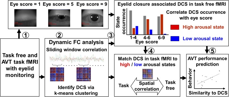
Fig. 1.
Graphic overview of the study. (Step 1) Task-free and AVT fMRI scans were collected following total sleep deprivation. The degree of SEC was rated. (Step 2) DFC analysis was performed to extract DCS. (Step 3) DCS corresponding to a high- and low-arousal state associated with eyelid closure were derived. (Step 4) DCS derived from the AVT task fMRI dataset were matched to their counterpart templates in the task-free condition. (Step 5) High- and low-arousal DCS thus derived had intersubject and within-subject behavioral correlates.

