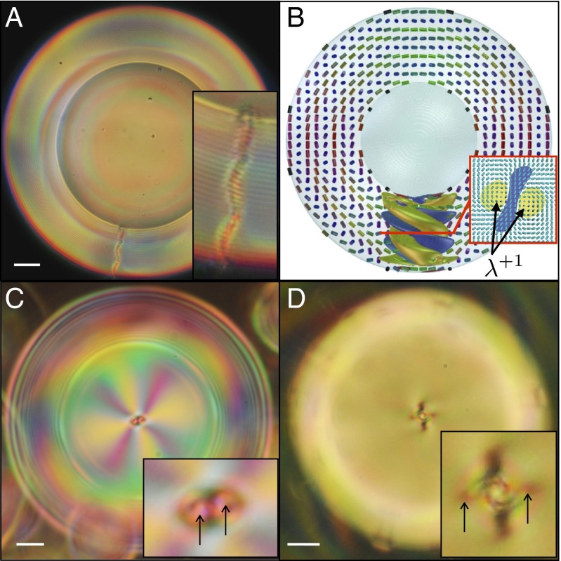
Fig. 3.
Cholesteric shells with monovalent defect structure. (A, C, and D) Cross-polarized images of experimental shells with different helical pitch p: 2.7, 3.5, and , respectively. (Scale bar, .) A is a side view of the shell, showing a radial defect spanning the shell, whereas B and C are top views of the shell, showing the two disclinations composing the radial defect. The arrows point to the ends of the disclinations, which wind around each other deeper down the shell. (B) Simulated cholesteric shell with radial spherical structure (RSS).

