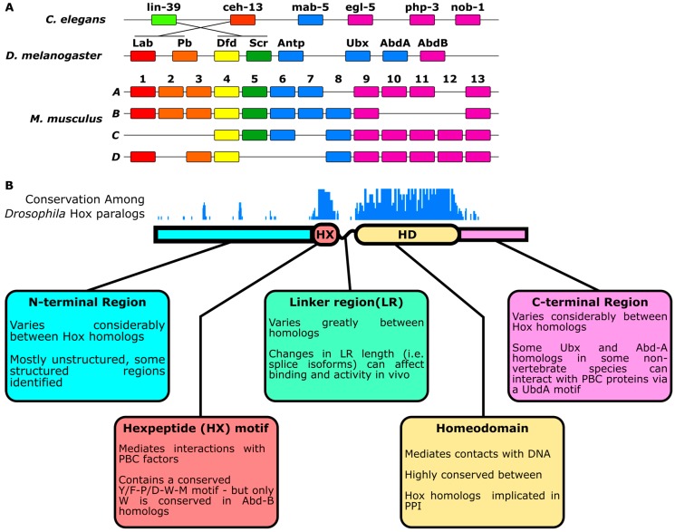
Figure 1.
Schematic of Hox gene locus and Hox proteins. (A) Schematic of Hox gene clusters in C. elegans, D. melanogaster and M. musculus. Genes with similar colors are thought to derive from a common ancestor [10]; (B) Schematic of a Hox protein with regions labeled and described in boxes (bottom) [11,12,13,14,15,16,17,18]. Per-amino-acid conservation score across Drosophila Hox protein sequences demonstrates that the HX and homeodomain regions are the most conserved regions across paralogous Hox proteins. Multiple sequence alignment was produced via ClustalΩ (top) [19,20]. Note: The size of each Hox protein region is not to scale.

