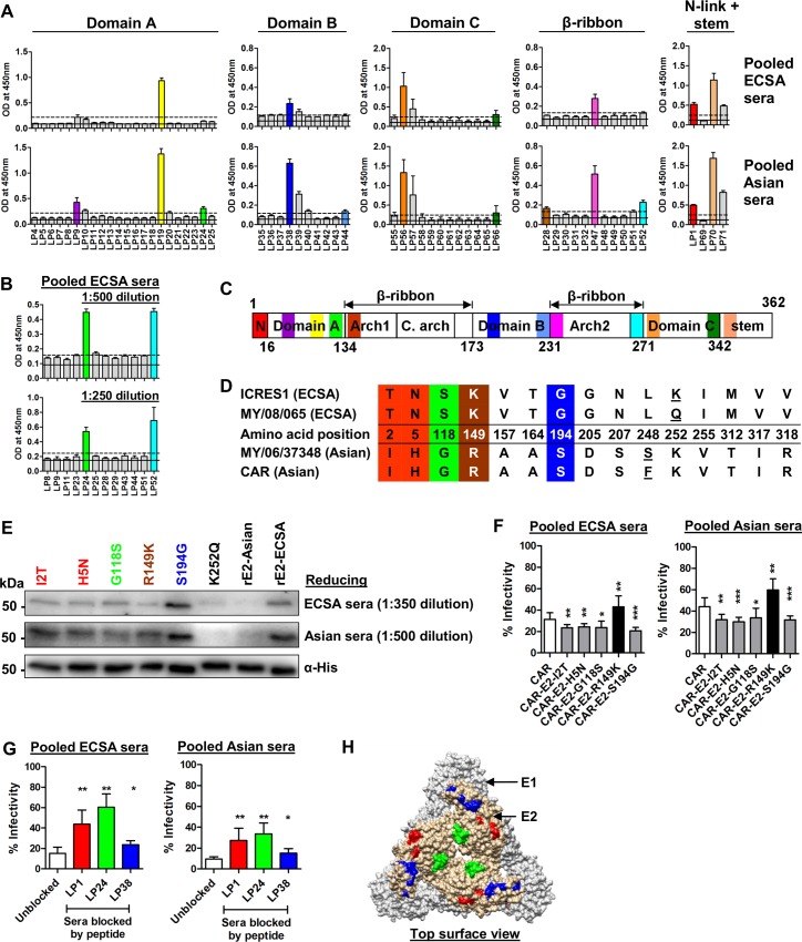
Fig 4. E2-I2T, H5N, G118S and S194G substitutions within linear neutralizing epitopes enhance the neutralization activity of ECSA and Asian sera.
(A) Overlapping synthetic peptides covering the E2 glycoprotein of MY/08/065 and its domains from amino acids 1–362 were screened with CHIKV immune sera at 1:1000 dilution. The black solid line represents the mean OD value of healthy controls and the dotted line represents the cut-off value (mean+3SD). The average results from 2 independent experiments are presented. In the event of two adjacent positively-mapping peptides, the peptide with the highest OD reading was taken. Key positive mapping peptides are color-coded. (B) Selected synthetic peptides were re-screened with pooled ECSA sera at lower dilutions of 1:500 and 1:250, in tetraplicate. The black solid line represents the mean OD value of healthy controls and the dotted line represents the cut-off value (mean+3SD). Key positive mapping peptides are color-coded. (C) Schematic diagram of the E2 protein showing the positions of the color-coded mapped epitopes. The numbers refer to the amino acid positions demarcating the E2 domains. N, N-link; C.arch, central arch. (D) Schematic representation of the E2 glycoprotein with the numbers indicating the amino acid positions of the glycoprotein and its domain proteins. The amino acid differences between E2 glycoproteins of ECSA (MY/08/065, ICRES1) and Asian (MY/06/37348, CAR) strains were tabulated and mapped (from amino acids 1–362). Amino acid differences within a genotype are underlined. Amino acid changes which fall within the identified linear epitopes are color-coded. (E) Immunoblotting was performed against recombinant E2 glycoproteins under reducing conditions, with each named amino acid change from the Asian to the ECSA sequence introduced independently. Mouse anti-His antibody was used as a control. (F) Seroneutralization was performed against different constructs with the CAR-2SG-ZsGreen backbone, which were rescued from the corresponding CHIKV icDNAs. Data are represented as means ± SD from 4 independent experiments at a serum dilution of 1:800 (pooled sera). *P<0.05, **P<0.01, ***P<0.001, Mann-Whitney U test. (G) Competitive peptide blocking assay was performed at 1:100 dilution with either pooled ECSA or Asian sera against ICRES1 at an MOI of 1. Data are expressed as percentages of infectivity of an infection control, and are presented as means ± SD from 2 independent experiments. *P<0.05, **P< 0.01, Mann-Whitney U test, relative to unblocked condition. (H) The color-coded mapped neutralizing epitopes are localized on the structural E1-E2 heterodimer complex (based on PDB 3J2W). The epitope sequence of LP1 is only partially localized as the 3D structure is not fully resolved.

