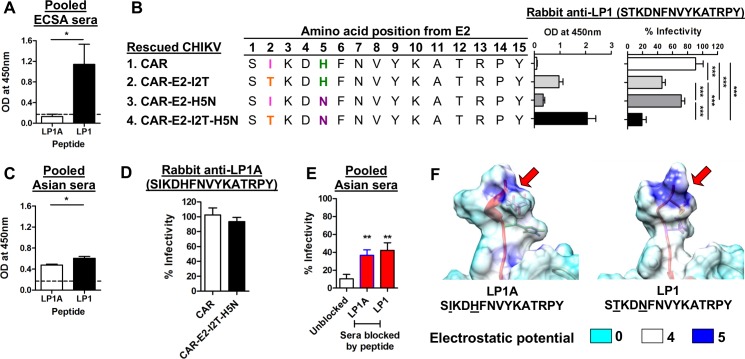
Fig 5. Sequence variation of a linear neutralizing epitope influences the spectrum of cross-neutralization across genotypes.
(A) Synthetic peptides LP1A and LP1 were screened with ECSA immune sera at 1:500 dilution in tetraplicate. The dotted line represents the cut-off value (mean + 3SD). *P<0.05, Mann-Whitney U test. (B) Recombinant viruses were pre-treated with 1% Triton X-100, then coated at 5 x 10⁵ pfu per well, and a binding assay was performed with 1μg/ml of antibody. The seroneutralization assay was performed with infection at an MOI of 50 against 25μg/ml of antibody. Data are represented as means ± SD from 3 independent experiments. ***P<0.001, repeated measures ANOVA with the Bonferroni multiple comparisons test. (C) Synthetic peptides LP1A and LP1 were screened with Asian immune sera at 1:500 dilution in tetraplicate. The dotted line represents the cut-off value (mean + 3SD). *P<0.05, Mann-Whitney U test. (D) Seroneutralization of anti-LP1A against CAR and CAR-E2-I2T-H5N at an antibody concentration of 25 μg/ml. Data are represented as means ± SD from 3 independent experiments.(E) Competitive peptide blocking assay was performed at 1:100 dilution with either LP1A or LP1 in pooled Asian sera against ICRES1 at an MOI of 1. Data are expressed as percentages of infectivity of an infection control, and are presented as means ± SD (n = 6). **P< 0.01, Mann-Whitney U test, relative to unblocked condition. (F) Structural images illustrating the changes of surface electrostatic potential due to differences in amino acid positions 2 and 5 within LP1A (Asian) and LP1 (ECSA) sequences (red arrows).

