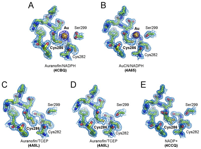
Figure 4. Cys286 gold-binding site in EhTrxR.
A–B, Gold atom originated from Auranofin (A) or AuCN (B) bound to Cys286. C–E, Free Cys286 adopts alternative conformations, only one of which is accessible for interactions with Au(I). Cys282 points away from Cys286 in all the structures resolved in this work. Fragments of 2Fo-Fc electron density map (blue mesh) contoured at 1.0 σ show gold binding site under different crystallization conditions. Au(I) is shown as a golden sphere, protein is drawn as sticks with heteroatoms colored according to the elements: oxygen in red, nitrogen in blue, and sulfur in yellow.

