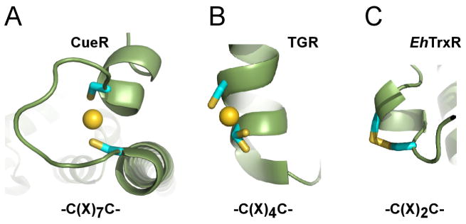
Figure 8. Dithiol C(X)nC motif.
A, A perfect geometry of the zeptomolar (10−21 M) chelate gold complex in CueR (Changela et al., 2003). B, C(X)4C motif of the high Mr thioredoxin-glutathione reductase (TGR) of S. mansoni (Angelucci et al., 2009) provides space to accommodate Au(I) but is too short to establish a two-coordinate linear configuration. Being in alternative conformations, one of the two cysteine residues separated by one α-helix turn, barely participates in gold binding. C, Spacing between the sulfhydryl groups in the C(X)2C motif of EhTrxR is insufficient to accommodate Au(I) and favors oxidation to disulfide instead.

