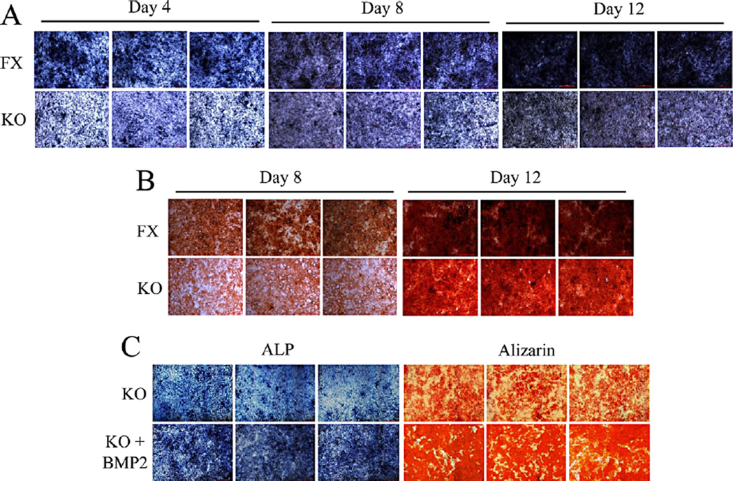
Fig. 5.
Deletion of Bmp2 delays cell differentiation and mineralization. A: The iBmp2fx/fx and iBmp2ko/ko dp cells were cultured in the calcifying medium for 4, 8, and 12 days. ALP activity was analyzed using in situ ALP staining. B: For cell mineralization assay, both of iBmp2fx/fx and iBmp2ko/ko dp cells were treated with calcifying medium for 8 and 12 days. Mineralized nodules were visualized with alizarin red S staining. C: The Bmp2ko/ko dp cells were treated either with or without recombinant Bmp2 (10 ng/ml) in calcifying medium for 7 and 14 days, respectively. The 7-day-inducced cells were used for ALP assay while 14-day induced cells were assayed by alizarin red S staining. Exogenous Bmp2 rescued the iBmp2ko/ko dp cell differentiation and mineralization. FX, floxed; KO, Bmp2 knock out.

