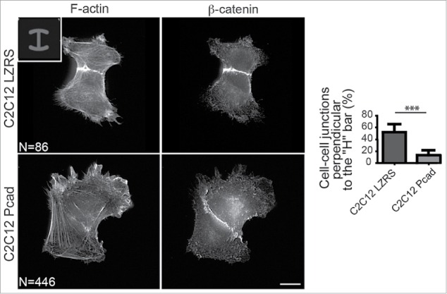
Figure 2.

P-cadherin expression decreases cell contractility as indicated by intercellular junction positioning. Immunostaining of β-catenin and F-actin in control (C2C12 LZRS: C2C12 cells expressing only the empty vector) and P-cadherin-expressing cell doublets plated on H-shaped micropatterns. Most of control C2C12 myoblasts (82%) have intercellular junctions that are perpendicular to the H bar. Conversely, the intercellular junction position and orientation are strongly perturbed upon P-cadherin expression. Indeed, junctions that are perpendicular to the H bar, like in control cells, are observed only in 11% of P-cadherin-expressing cells. For all panels, the mean ± SEM is shown; *** P < 0.0005.
