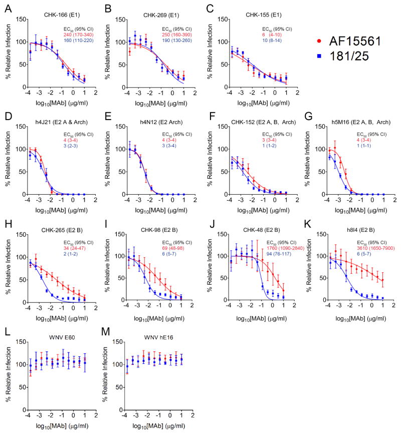
Figure 7. Differential Neutralization of AF15561 and 181/25 by MAbs Targeting E2 Domain B.
(A–M) MAbs were incubated with 100 FFU of AF15561 (red) or 181/25 (blue) viruses for at 37°C for 1 h. MAb-virus mixtures were added to Vero cells and incubated for 18 h. Virus-infected foci were stained and counted. Wells containing MAbs were compared with wells without MAbs to determine relative infection. (L) WNV E60 and (M) hE16 MAbs were included as isotype control MAbs. The concentrations at which 50% of AF15561 (red text) or 181/25 (blue text) were neutralized (EC50) were determined by non-linear regression and are displayed in ng/ml (95% CI). Each graph represents the mean and standard deviation (SD) from at least two independent experiments.

