Figure 3.

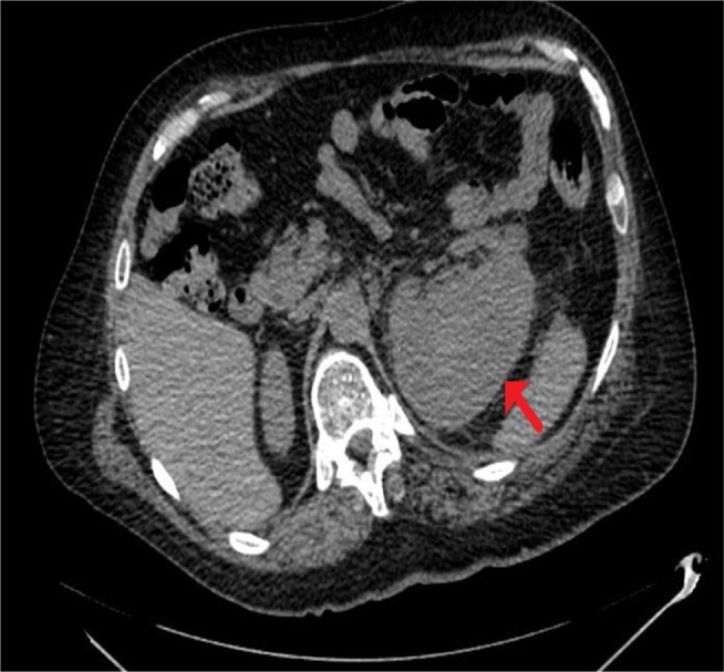
CT of urinary tract.
Note: Performed 4 weeks after the third ureteroscopy, axial section: 9×7 cm subcapsular collection on the left kidney compressing the underlying parenchyma present (arrow).
Abbreviation: CT, computed tomography.

CT of urinary tract.
Note: Performed 4 weeks after the third ureteroscopy, axial section: 9×7 cm subcapsular collection on the left kidney compressing the underlying parenchyma present (arrow).
Abbreviation: CT, computed tomography.