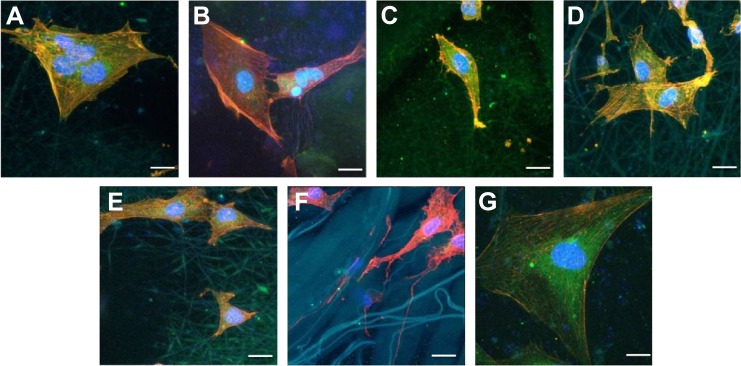
Figure 6.
CLSM (×400) of morphology of cells cultured on five different membranes compared with untreated coverslips and Bio-Gide Collagen membranes.
Notes: (A) PLGA/PCL ES. (B) PLGA/PCL. (C) PLGA/PCL/COL I ES. (D) pDA–PLGA/PCL ES. (E) COL I–pDA–PLGA/PCL ES. (F) Bio-Gide Collagen membranes. (G) Untreated coverslips. Scale bars =20 μm. Cells were stained for F-actin (red), nucleus (blue), and substrates (green) by fluorescent dyes. Cell adhesion on the five different membranes was demonstrated by cellular morphology at 12 hours after seeding onto the membranes compared to untreated coverslips and Bio-Gide Collagen membranes.
Abbreviations: CLSM, confocal laser scanning microscopy; PLGA, poly(lactic-co-glycolic acid); PCL, poly(caprolactone); ES, electrospinning; COL I, collagen I; pDA, 3,4-dihydroxyphenylalanine.

