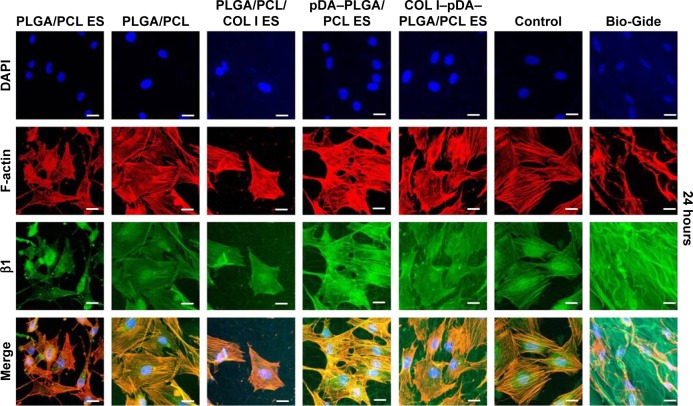
Figure 9.
Localization of β1 integrin and F-actin protein was identified by immunofluorescence staining at 24 hours after seeding on five different membranes compared with untreated coverslips (control) and Bio-Gide Collagen membranes (PLGA/PCL ES; PLGA/PCL; PLGA/PCL/COL I ES; pDA–PLGA/PCL ES; COL I–pDA–PLGA/PCL ES).
Notes: Cells were observed by CLSM (×400) and immunostained for F-actin (red), nucleus (blue), and β1 (green). The bottom rows in each panel show the merged images. Control, untreated coverslips; Bio-Gide, collagen membranes. Scale bars =20 μm.
Abbreviations: PLGA, poly(lactic-co-glycolic acid); PCL, poly(caprolactone); ES, electrospinning; COL I, collagen I; pDA, 3,4-dihydroxyphenylalanine; CLSM, confocal laser scanning microscopy; DAPI, 4′,6-diamidino-2-phenylindole.

