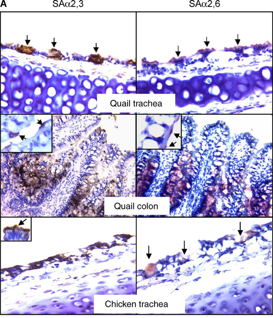
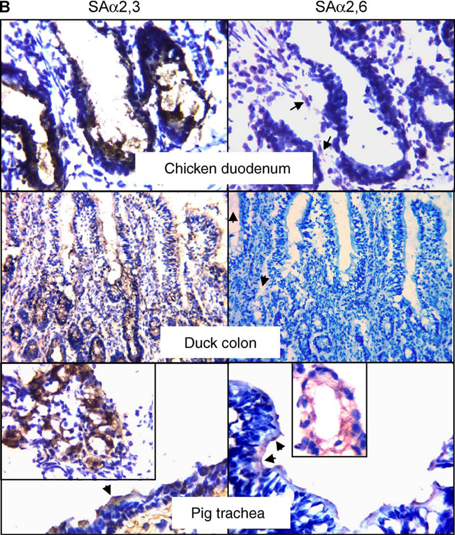
Fig. 1.
Immunologic detection of SAα2,3-gal and SAα2,6-gal linked receptors in quail, chicken, duck and pig. Sections were exposed to specific agglutinins and corresponding antibody for immunostaining and were counterstained with hematoxylin. The brown color (developed from DAB) indicates the presence of SAα2,3-gal. The red color (developed from AEC) highlights the presence of SAα2,6-gal. The original magnification of quail colon and duck colon is 400×, while those of the remaining sections are at 1000×. Insets are magnified views of SAα2,3-gal or SAα2,6-gal present in epithelial cells of corresponding tissues, while those in pig photos show the staining of receptors in seromucous glands. Arrows emphasize the positively stained cells. Note that, in photos of quail trachea, the staining for SAα2,3-gal is located mainly in mucin-producing cells, whereas the staining for SAα2,6-gal is seen mainly in ciliated cells. The arrows in the photo of chicken duodenum show slight, nonspecific staining in the connective tissue. Likewise, weak positive for SAα2,6-gal staining is detected in the connective tissue of duck intestine.


