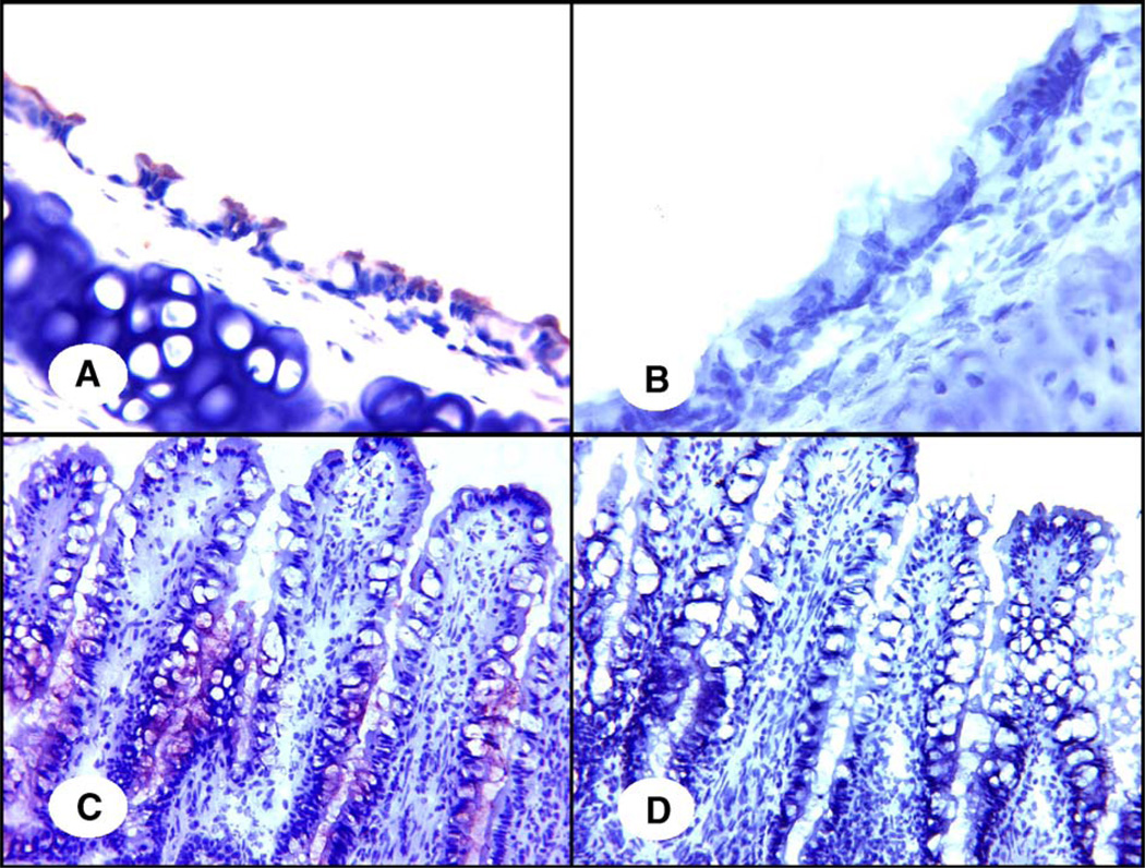
Fig. 2.
Specificity of the reaction between SNA and quail tissues. In panels A and C, the SAα2,6-gal-specific SNA (1 µg/ml) was applied directly in the lectin staining as described under Materials and methods. In panels B and D, the SAα2,6-gal specific SNA was incubated with 20 µg/ml transferrin before staining. Panels A and B indicate the trachea; panels C and D indicate the colon. Original magnification is 400×. Red staining indicates the presence of SAα2,6-gal receptors.

