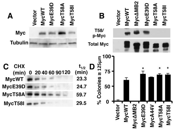
Figure 1. Burkitt’s Lymphoma Myc mutants enhance Myc-induced cell transformation independently of loss of T58 phosphorylation and increased expression.
A) Rat1a fibroblasts were engineered to express MycWT, the indicated Burkitt’s Lymphoma (BL) Myc mutants or vector control. Cell extracts were analyzed by Western blot using anti-Myc or anti-Tubulin antibodies. B) MycWT and the BL-associated Myc mutants were transiently expressed in 293 cells. Cell extracts were analyzed by parallel western blots using anti-phospho-Myc (p58/p62) or anti-Myc (phospho-independent). C) The half-life of MycWT and BL-associated mutants was determined by engineering myc−/− fibroblast to express the indicated proteins, and cells were treated with 50 μg/mL cycloheximide for the up to 120 minutes. Cell extracts were analyzed by western blot using anti-Myc antibodies D) Log phase Rat1a fibroblasts expressing MycWT or BL-associated Myc mutants were used in a soft agar transformation assay. The chart depicts the diameter of colonies from each cell line after 1 week of growth. Error bars indicate the standard deviation (SD) for three biological replicates using two independently engineered cell line. Significance was determined using student’s t-test; *p≤0.05.

