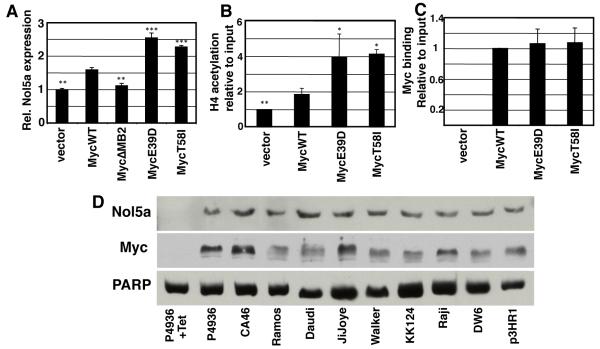
Figure 4. The nucleolar protein gene, Nol5a, is differentially induced and acetylated by MycE39D and MycT58I, and highly expressed in Burkitt’s Lymphoma cell lines.
A) The relative expression level of Nol5a in Rat1A fibroblasts engineered to express MycWT or the indicated BL associated mutations was determined by qPCR. B) ChIP assays were performed at the same site in the Nol5a promoter using anti-acetylated H4 antibodies to determine relative acetylation by the indicated myc proteins. C) ChIP assays were performed using anti-Myc antibodies to determine Myc binding at a conical E box in the Nol5a promoter. D) P493-6 human B cells express high levels of Myc which can be repressed by tetracycline. After 72 hour treatment with 0.1 μg/mL tetracycline or vehicle, nuclear lysates for P493-6 cells as well as indicated Burkitt’s Lymphoma cell lines were prepared and analyzed by western blots using an anti-c-myc and anti-Nop56 antibodies. Error bars (SD) represent three biological replicates. Asterisk indicates significant difference compared to MycWT; student’s t-test, *p≤0.05, **p≤0.01,***p≤0.001.

