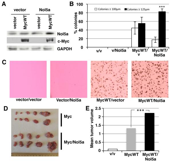
Figure 6. Nol5a synergizes with Myc WT to increase cellular transformation and tumor size.
A) Rat1A fibroblast were engineered to express control vector, Nol5a, and/or MycWT. Lysates were assayed by western blot using anti-Nol5a (Sigma), anti-Myc, and anti-GAPDH. B) Cells expressing the indicated vectors were plated in soft agar and cellular transformation was measured by colonies formation and scored for size after 7 days. Colonies were scored as ≤ 100 μm or ≥ 125 μm divisions using a reticule; student’s t-test, p≤0.001 C) Representative soft agar plates expressing indicated vectors 7 days after plating. D) Rat1A cells engineered to express vector (n=10), MycWT (n=10), or MycWT/Nol5a (n=10) were injected subcutaneously into the flanks of nude mice and allowed to form tumors. The tumors were extracted after 3 wks of growth. E) Individual tumors were weighed and mean mass for each tumor type was calculated. The mass of tumors induced by MycWT, and MycWT/Nol5a were determined to be significantly different; student’s t-test, p≤0.001.

