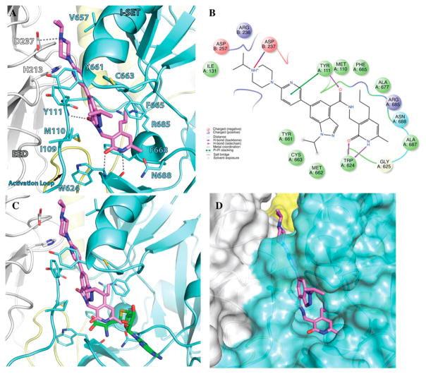
Figure 3.
Docked pose of compound 5 in the human PRC2 structure. (A) Docked pose of compound 5 (pink). (B) A 2D representation of molecular interactions between compound 5 and the residues of the binding site. (C) Superimposition of compound 5 and SAH (aligned from PDB entry 5HYN). The pyridone moiety of compound 5 mimics the homocysteine moiety of SAH (green). (D) Binding site of PRC2 complex where compound 5 binds through a groove formed by the SET domain and the activation loop of EZH2 (aqua) and the EED complex (gray).

