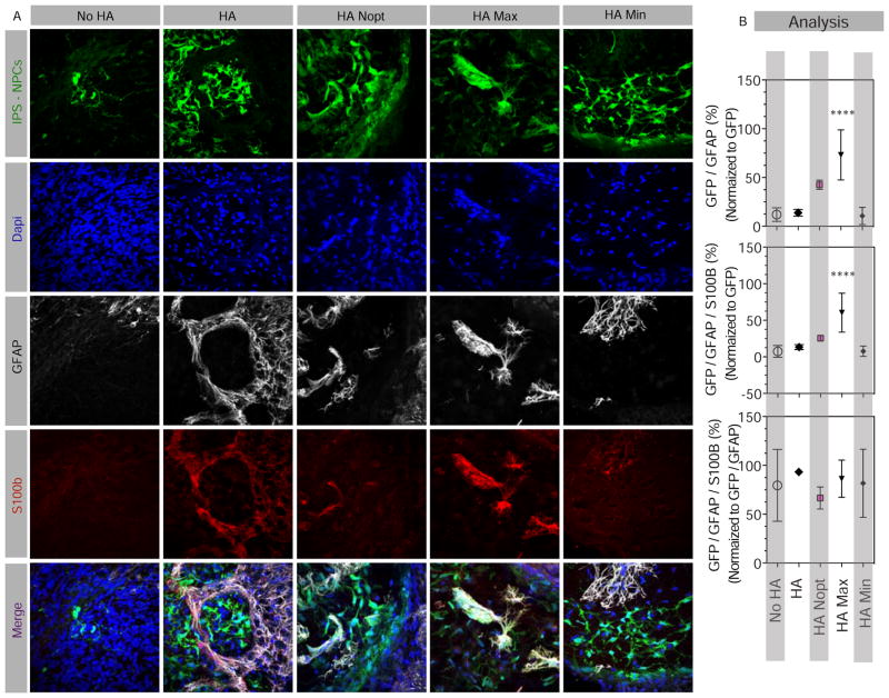
Figure 4. Effect of HA Max composition on astrocytic cell differentiation.
(A) Fluorescent images and (B) analyses of brain sections injected with 100,000 cells stained at 6 weeks for GFP, GFAP, as the marker for both astrocytes and NPCs, and S100b that is more specific to astrocytes. The numbers of GFP/GFAP as well as GFP/GFAP/S100b positive cells were normalized to the total GFP cells to obtain a percentage cells expressing one or both astrocytic markers. In addition, triple labeled cells were normalized to the number of GFP-GFAP cells in order to estimate the percentage of GFAP cells that are also positive for the more exclusive astrocytic marker, S100b. Scale bars, 50 μm. **** indicates P < 0.0001.

