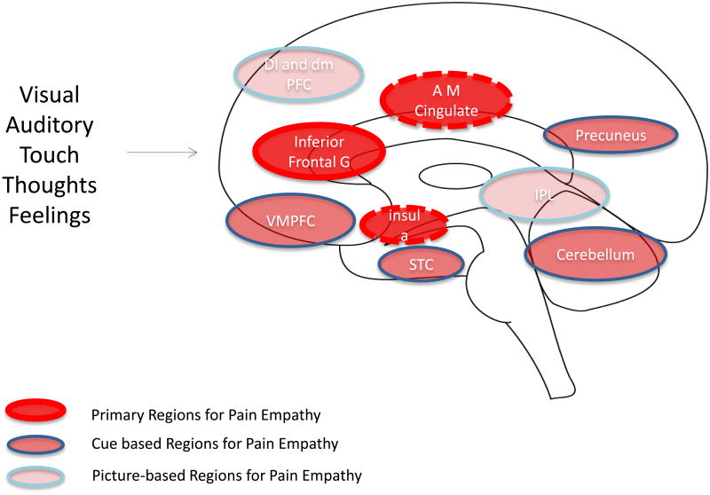
Figure 2. Key Brain Regions for Empathy.
The figure (adapted from (Lamm et al., 2011) summarizes data implicating complex neural networks that overlap for empathy: These include (1) Primary Regions for Pain Empathy which themselves have a large overlap with pain activations in experimental pain; (2) Cue based Regions for Pain Empathy that encompass complex visual, social and other signals; and (3) Picture-based Regions for Pain Empathy that aside from the specific salience of pictures used in the experimental setting, clearly visual inputs related to pictures may contribute to an aversive process. Although only one side of the brain is depicted in the figure, these results are not lateralized.

