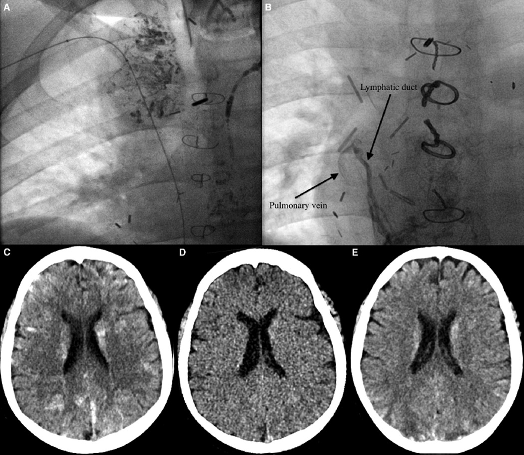
Figure 1.
A, Chest radiograph demonstrating lipiodol in peribronchial lymphatic ducts. B, Chest radiograph demonstrating an abnormal connection between a lymphatic duct and the pulmonary vein. C, Noncontrast CT shows numerous hyperdensities scattered throughout the brain bilaterally. D, A virtual noncontrast CT image derived from dual energy CT proves that the hyperdensities were iodine containing material (lipiodol) rather than just multifocal hemorrhage. E, CT image from 5 days later shows mild interval decrease and improvement in the hyperdensities, with development of foci of hypodensity (eg, in the bilateral posterior parietal lobes).

