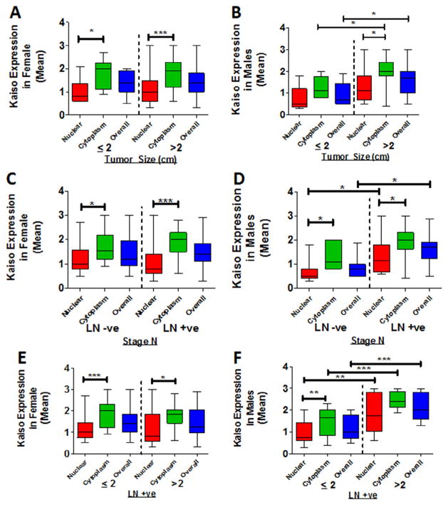
Fig. 3.
Cytoplasmic and nuclear expression of Kaiso in male and female PDAC patients. Cytoplasmic and nuclear expression of Kaiso was compared in tumors ≤2 or ≥2 cm in size. (A) There were no significant differences in cytoplasmic or nuclear Kaiso expression in tumors of females. (B) Tumors of males ≥2 cm in size demonstrated higher levels of cytoplasmic (P = 0.0384) and nuclear Kaiso (P = 0.0236). (C) Tumors of females did not demonstrate a significant difference with positive or negative lymph nodes (Stage N). (D) For lymph node-positive patients compared to lymph node-negative patients, tumors of males showed elevated Kaiso in the nucleus (P = 0.0413) and overall (P = 0.0141). (E) For females, there was no significant difference in tumors ≥2 or ≤2 cm in size that had positive or negative lymph nodes. (F) Tumors of males ≥2 cm in size with positive lymph nodes showed elevated expression of Kaiso in the cytoplasm (P = 0.0001), in the nucleus (P = 0.0012), and overall (P = 0.0002). LN+ve, lymph node positive; LN−ve, lymph node negative. *P < 0.05 is significant. **P < 0.01 is significant. ***P < 0.001 is significant.

