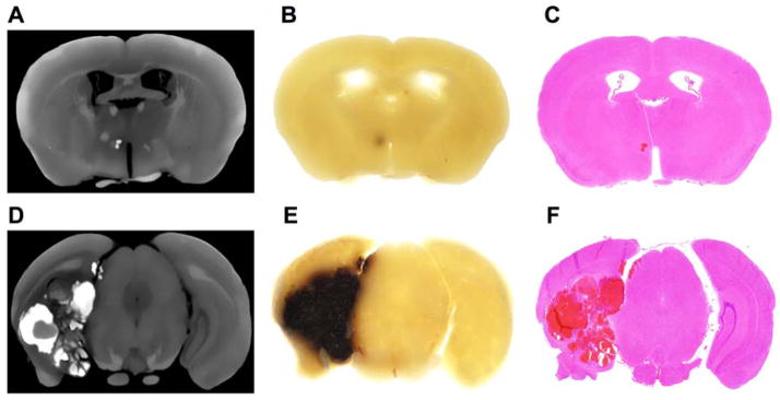
Fig. 2.
Identification of small and large CCM lesions in a transgenic murine model of cerebral cavernous malformation. (A, D) Coronal section of a micro-CT generated image showing a hyper-dense stage 2 CCM lesion. (B, E) Following micro-CT acquisition, the corresponding gross slice (1-mm thickness) was examined at 1.5x magnification using a dissecting microscope (Olympus SZ61 mounted with an Olympus DP21 camera) to assess the presence of CCM lesions. (C, F) The hyper-dense structure was validated to be a CCM lesion using histology. (D–F) Illustration of a Ccm3+/−Trp53−/− murine model (not included in our analysis) highlighting the difficulty of assessing lesion burden in large complex stage 2 CCM lesions on single cross sectional histologic section. This limitation is overcome by using volumetric micro-CT analysis of lesion burden.

