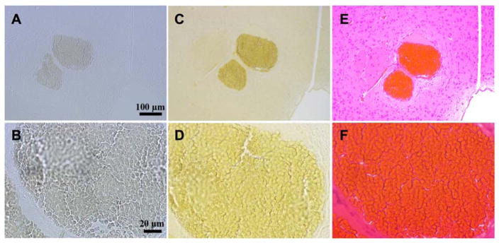
Fig. 4.
Iodine in blood-filled caverns of CCM lesions. (A) 10X and (B) 40X magnification of unstained 5-μm histological sections showing the CCM lesion. (C) 10X and (D) 40X magnification of an iodine-stained section demonstrating the higher concentration of iodine in the blood-filled cavern relative to surrounding parenchyma. (E) 10X and (F) 40X magnification of the H&E stained section of the corresponding lesion showing the morphology of the blood filled cavern with surrounding parenchyma.

