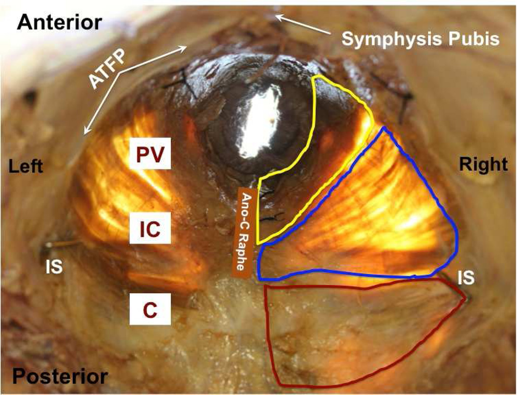
Figure 1.
Representative photograph of in situ pelvic floor muscles from a younger donor (superior view) with superimposed boundaries of coccygeus (C, red), iliococcygeus (IC, blue), and pubovisceralis (PV, yellow).
ATFP: arcus tendinious fascia pelvis; IS: ischial spine; Ano-C Raphe: ano-coccygeal raphe.

