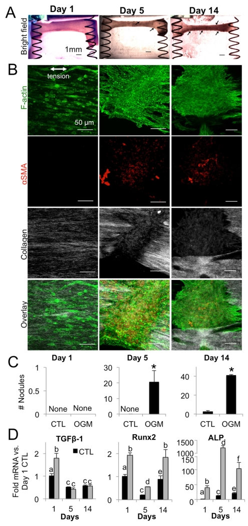
Fig. 7. Nodules and osteogenic signaling in mechanically active VIC hydrogels.
A. Bright-field images of osteogenic hydrogels at day 1, 5, and 14. Arrows indicate nodules. B. Immunofluorescence of osteogenic hydrogels at day 1, 5, and 14. Green = f-actin, red = αSMA, white = collagen. C. Number of nodules formed in control and osteogenic hydrogels at day 1, 5, and 14. D. TGF-β1, Runx2, and ALP mRNA in control and osteogenic hydrogels at day 1, 5, and 14. N =4 for all experiments. Bars with different letters are statistically different (p< 0.05), error bars indicate SEM.

