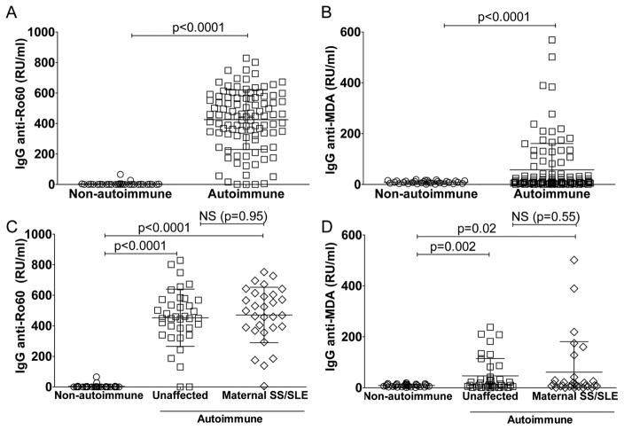
Figure 7. Detection of maternal IgG autoantibodies in cord blood.
A–B. Levels of maternally transferred IgG-autoantibodies to Ro60 or the oxidation-associated malondialdehyde (MDA) in 31 control cord bloods (non-autoimmune) or 103 cord blood samples from the neonatal registry cohort of mothers with IgG anti-Ro/La autoantibodies (autoimmune). C–D. Levels of IgG-autoantibodies to Ro60 and MDA in 31 control cord blood samples (non-autoimmune), compared to 33 cord blood samples of newborns with mothers enrolled in the NL cohort but without documented signs and/or symptoms or prior diagnosis of autoimmune disease at the time of birth (unaffected), or 30 newborns from mothers with a preceding history of systemic lupus erythematosus (SLE), primary Sjögren’s syndrome (SS), or both (SS/SLE). P-values were derived two-sided unpaired t-test with Welch’s correction.

