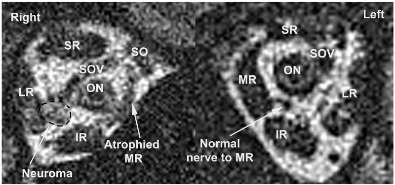
FIG 4.
Contrast-enhanced coronal T1-weighted surface coil MRI of right orbit of case 2 demonstrating a 3.3×2.8 mm enhancing lesion in the path of the motor nerve to the medial rectus muscle compatible with schwannoma. IR, inferior rectus muscle; LR, lateral rectus muscle; MR, medial rectus muscle; ON, optic nerve; SOV, superior ophthalmic vein; SR, superior rectus muscle.

