
Figure 2. ICV injection of AAV2/9-TDP-43 leads to expression of TDP-43 protein throughout mouse brain.
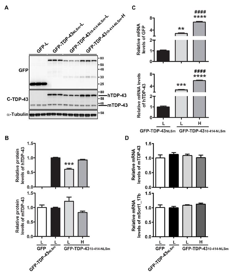
Western blot (A) and densitometric quantification analysis (B) revealed that the levels of GFP-TDP-4310-414-NLSm protein at high dose were comparable to that of GFP-TDP-43NLSm at low dose. Expression of GFP-TDP-43NLSm and GFP-TDP-4310-414-NLSm did not reduce the protein levels of endogenous mouse TDP-43 (B). (C) Quantitative RT-PCR (qRT-PCR) using specific primers against GFP or human TDP-43 showed that the mRNA levels of GFP-TDP-4310-414-NLS were dramatically higher than that of GFP-TDP-43NLS. (D) qRT-PCR using specific primers for mouse TDP-43 or mouse sortin1_17b showed that expression of GFP-TDP-43NLS or GFP-TDP-4310-414-NLS did not reduce mRNA levels of mouse TDP-43 nor induce aberrant splicing of mouse sortilin 1. Data shown are the means ± SEM of 3 mice per group. ** P<0.01, *** P<0.001, **** P<0.0001 versus GFP-TDP-43NLSm (L), #### P<0.0001 versus GFP-TDP-4310-414-NLSm (L), as assessed by one-way ANOVA with Tukey’s post-hoc analysis.