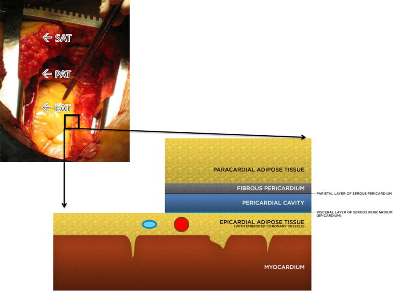
Figure 1. Epicardial adipose tissue (EAT) is an anatomically distinct adipose depot.

EAT lies on the myocardial surface without fascial separation such that epicardial adipocytes are in direct contact with the myocardium, even appearing to invaginate the myocardial surface on histology. In healthy adult subjects, EAT lies in the atrioventricular grooves and follows the branches of the major coronary arteries such that the coronary vessels are embedded within the EAT, although some patients may present with a massive epicardial fat pad as shown. Superficial to the epicardial fat are the visceral and parietal layers of the pericardial sac. Paracardial adipose tissue (PAT) is superficial to the pericardium and thus has no direct anatomical contact with the EAT or myocardium; it also has a separate microcirculation from EAT, as EAT shares the microcirculation of the heart. Paracardial fat is alternatively termed mediastinal fat. Although EAT and PAT are anatomically distinct, they are sometimes discussed under the umbrella term pericardial fat which refers to epicardial plus paracardial fat (6).
