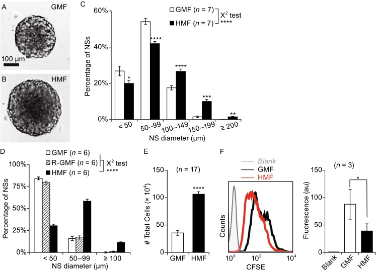
Figure 1.

Exposure to the HMF accelerates the growth of primary NSs from neonatal mouse. Primary cells from whole brains of P2 mice were seeded in either 60 mm dishes (8.0 × 10⁵ cells/dish for cell counting) or 96-well plates (1000 cells/well for NS counting and size analysis) and exposed to either the GMF or HMF. (A and B) Representative pictures of NSs at day 7. Those grown in the HMF appeared significantly larger. (C) Size distribution of day 7 NSs. A greater number of large NSs were counted in cultures exposed to the HMF. (D) Size distribution of day 6 NSs cultured in the GMF, R-GMF, and HMF conditions. Sizes of NSs in the GMF and R-GMF groups were similar, but smaller than those in the HMF group. (E) Total cell numbers of the day 7 NS cultures were significantly greater in the HMF group, compared to the control GMF group. (F) Cells exposed to the HMF underwent more divisions, as shown by the significantly decreased CFSE fluorescence and lower mean fluorescence. Data are shown as mean ± SEM (C and E) or SD (F). n is the number of animals (C–E) and the trials (F) used in the experiments. The P-value was calculated using a χ2 test for NS size distributions in (C) and (D), a one-way ANOVA for mean comparisons in (E). The two-tailed paired student’s t-test in (F) *P < 0.05; **P < 0.01; ***P < 0.001; ****P < 0.0001; n.s. P ≥ 0.05
