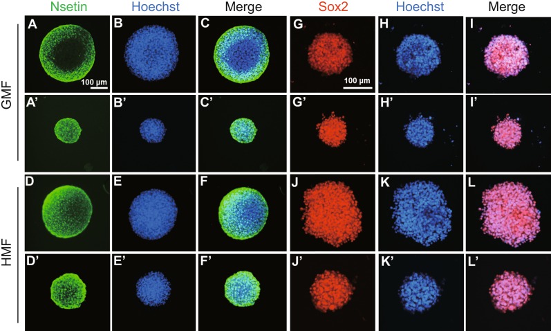
Figure 3.
The HMF-exposed NSs were positive of nestin and Sox2. Primary cultures of day 7 NSs from P2 mice were collected and immunostained with the neural stem cell markers, nestin (green) and Sox2 (red). Nuclei are stained with Hoechst (blue). Panels show representative large (A–L) and medium-sized (A’ –L’) NSs from the GMF (A–C/A’–C’; G–I/G’–I’) and HMF (D–F/D’–F’; J–L/J’–L’). The NSs of different sizes from both GMF and HMF condition were positive for nestin and Sox2

