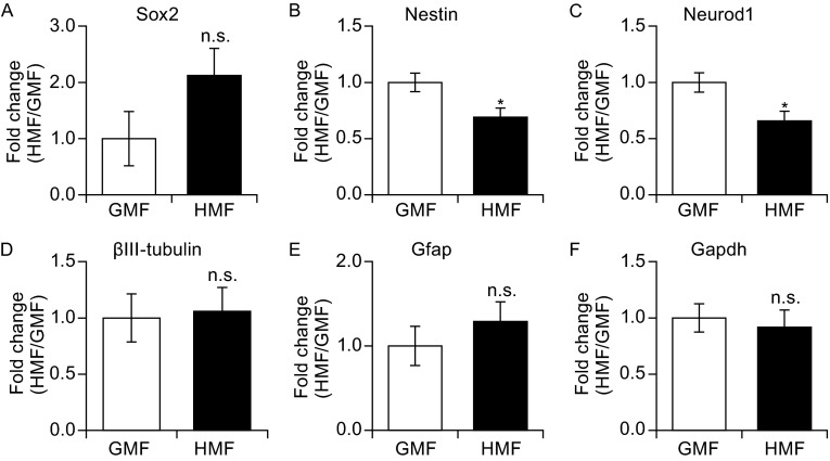
Figure 4.
The HMF-exposed NSs showed altered genes expressions. When checked expressions of the NSCs markers (Nestin; Sox2), differentiated neuronal markers (Neurod1; ßIII-tubulin) and glial marker (Gfap), the expression of both Nestin (B) and Neurod1 (C) were significantly down-regulated following exposure to the HMF. Compared to the GMF groups, the Sox2 had a trend of increase in the HMF group (A).The ßIII-tubulin and Gfap were found no significant changes (D and E). Tubulin 5α was used as the internal reference gene, and Gapdh was used as another internal reference gene (F). Data are shown as mean ± SEM from three independent experiments, six animals per expeiment. P values were calculated by one-way ANOVA for mean comparisons. *P < 0.05; n.s. P ≥ 0.05

