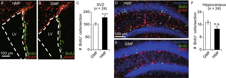
Figure 8.

HMF exposure promotes cell proliferation in the adult mouse brain. Adult male mice (4 to 6-week-old) were reared in either the HMF or GMF conditions for 30 days. Representative images of the SVZ are shown in panels (A) and (B), and the hippocampus in (D) and (E). Proliferative cells were immunostained with anti-BrdU antibody (green) and glia cells with anti-GFAP antibody (red). Nuclei were counterstained with Hoechst (blue). White dash lines outline the edge of the lateral ventricles (LV). (C and F) show number of BrdU-positive cells per section from the SVZ (C) and hippocampus (F). Exposure to the HMF increased the number of proliferative cells in the SVZ but not the hippocampus. n indicates the number of animals from at least three independent experiments. Data are shown as mean ± SEM. P values were calculated using a one-way ANOVA for mean comparisons. ****P < 0.0001; n.s. P ≥ 0.05
