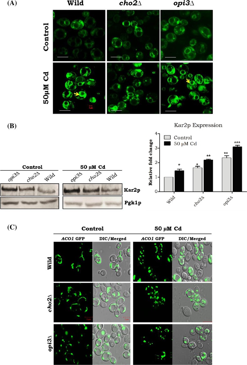
Fig. 5.
Defects in PC methylation in inducing membrane proliferation, ER stress, and mitochondrial damage. Wild-type and mutant cells were grown up to mid-log phase on YPD medium with or without Cd at 30 °C. a Membrane proliferation was assessed by the incorporation of the lipophilic dye DiOC6 and viewed under the fluorescence microscope. The bar in the figures indicates the scale of 1.0 μm. b Cells were lysed, and 50 μg of protein was loaded and separated by 10 % SDS-PAGE followed by immunoblotting with kar2 antiserum. Band intensity was quantified by the ImageJ software and expressed as a ratio with pgk1. c The ACO1-pGFP was analyzed for mitochondria morphology. Strains wild-type and cho2∆ contain typical tubular mitochondria. Opi3∆ caused mitochondrial fragmentation gradually increased; Aco1p showed 100 % mitochondrial network fragmentation in all Cd-treated mutant yeast cells

