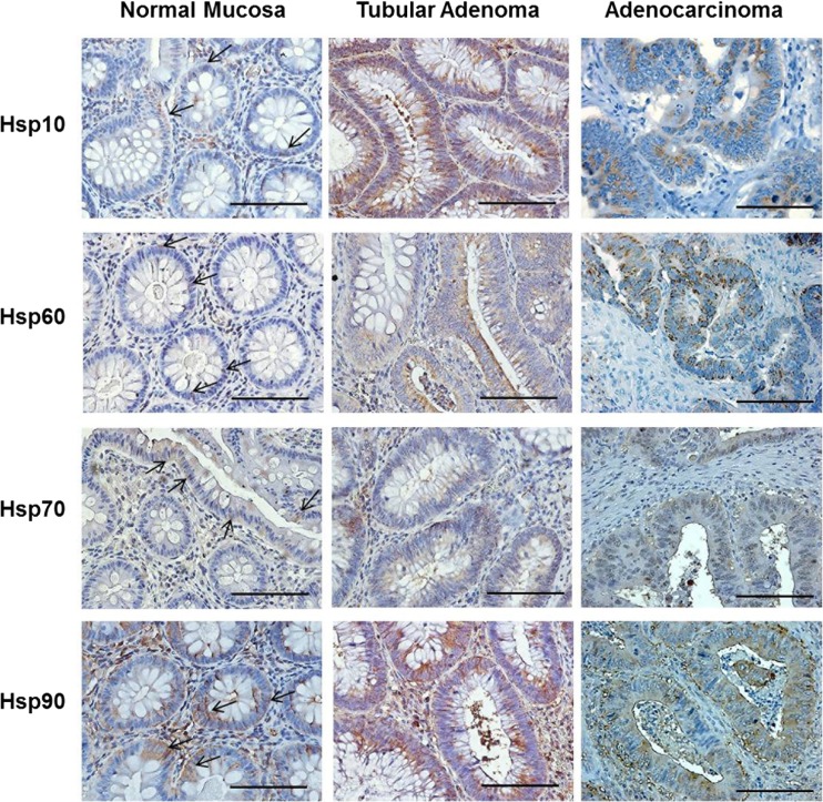
Fig. 2.
Representative images of immunohistochemical results for Hsp10, Hsp60, Hsp70, and Hsp90 in human large bowel biopsies of normal mucosa, tubular adenoma with moderate grade of dysplasia, and adenocarcinoma with moderate grade of differentiation. Magnification 400×. Scale bar 100 μm. Arrows show the positivity for the Hsps in the epithelial cells of normal mucosa. Note the slight positivity for Hsp60 in normal epithelial cells

