Figure 1.
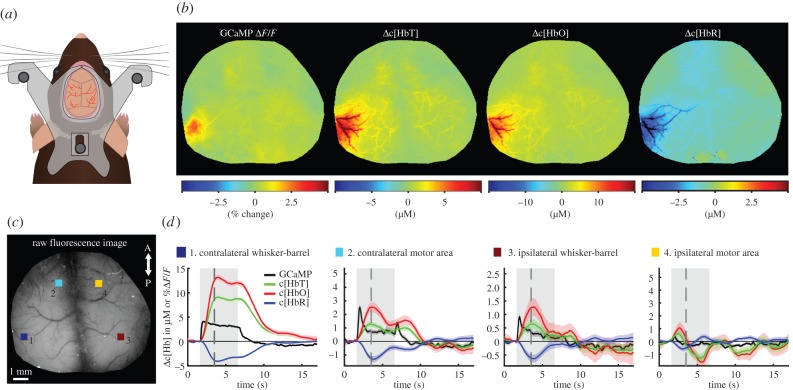
Demonstration of WFOM imaging of neural activity and haemodynamics in awake, behaving mouse brain. (a) A custom-made acrylic head plate is surgically implanted onto a thinned skull cranial window. After recovery, the mouse's head is held by attaching the head plate to an aluminium head plate holder to restrict head motion during imaging. The animal is free to run or rest on a saucer wheel positioned below. (b) Functional maps of Thy1-GCaMP6f (selectively expressed in excitatory neurons of layers 2/3 and 5 [17]) ΔF/F after haemodynamic correction, Δ[HbT], Δ[HbO] and Δ[HbR] 1.7 s after stimulus onset. (c) Grey scale raw fluorescence image of the cortical surface with four selected regions of interest (ROIs) in the contralateral (1) whisker-barrel and (2) motor areas, as well as in the ipsilateral (3) whisker-barrel and (4) motor areas. (d) Time course of neural GCaMP neural activity and haemodynamic changes in HbT, HbO and HbR corresponding to these four regions. Grey shading shows stimulus time. Black dashed line shows time of frames shown above. Data were acquired using Andor Ixon camera with 512 × 512 frames and 30 ms exposure time per frame. Data shown are an average of 38 repeated stimulation trials in one mouse, with 5 s duration, 25 Hz tactile whisker stimulation.

