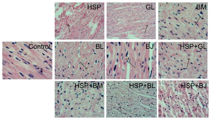
Figure 2.
Histopathological results of the heart by H&E staining. Control group: no necrotic lesions; HSP: heishunpian group, infiltration of lymphocytes; GL: gualou group, irregular arrangement of cell nuclear; BM: beimu group, inhomogeneous staining of cytoplasm; BL: bailian group, cytoplasm discoloration and uneven staining; BJ: baiji group, overflow of red blood cells in muscle fibers; HSP + GL: heishunpian-gualou group, uneven staining of cytoplasm; HSP + BM: heishunpian-beimu group, mild hyperchromatic of cell nuclear; HSP + BL: heishunpian-bailian group, loss of muscle fibers and irregular arrangement of cell nuclear; HSP + BJ: heishunpian-baiji group, atrophy of striated muscle.

