Figure 6.
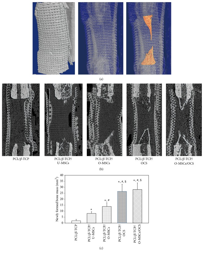
Bone regeneration in canine radial defects. (a) 3D reconstructed image and (b) sagittal view image showed that new bone formation was detected within defects at the bone margin. (c) Quantitative 3D micro-CT analysis revealed that groups with cell sheets (with or without O-MSCs) showed a greater volume of newly formed bone than the other groups (∗, #, $ P < 0.05). ∗: compared to the PCL/β-TCP group, #: compared to the PCL/β-TCP/U-MSCs group, and $: compared to the PCL/β-TCP/O-MSCs group.

