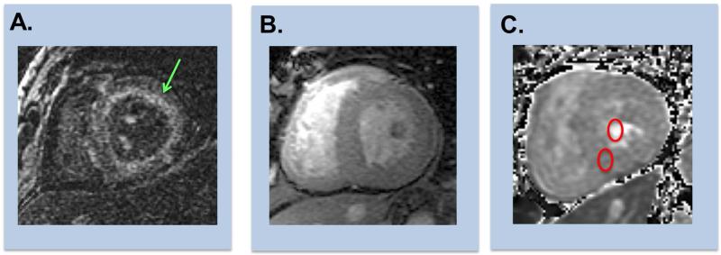
Figure 4. Representative Examples of Cardiac Amyloid on CMR.
Representative examples of CMR evidenced enhancement patterns among patients with cardiac amyloid.
(A) Diffuse subendocardial enhancement (green arrow) on DE-CMR (inversion time [TI] 300msec).
(B) Diffuse transmural enhancement on DE-CMR (left [equivalent TI]).
(C) Corresponding T1 map enables quantification of extracellular volume fraction, which can be can be calculated via measurement of T1 in myocardium and blood pool (red circles) on matched pre- and post-contrast images.

