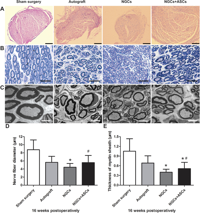
Figure 8. Histological assessment of the regenerated sciatic nerves in the distal segment at 16 weeks after surgery.
(A) H&E staining shown the overview of nerve morphology in each group (Scale bars = 200 μm). Myelination of regenerated nerves revealed by toluidine blue staining (B, scale bars = 25 μm) and TEM (C, scale bars = 5 μm). Statistical analysis of the diameters of myelinated nerves (D) and thickness of myelin sheath (E) for each group (n = 4). ∗p < 0.05 for comparison with autograft group, and #p < 0.05 for comparison with NGCs group.

