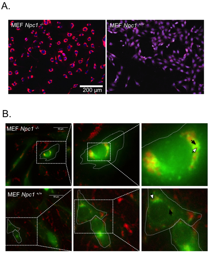
Figure 1. Npc1 deficiency leads to cholesterol accumulation in enlarged late endosomes/lysosomes.
(A) Filipin staining was performed to visualize subcellular cholesterol in Npc1−/− and Npc1+/+ MEFs. A high throughput microscope was used to compare subcellular cholesterol (psuedo colored red) in both cell types. Nucleus was stained using HCS nuclear mask deep red stain. (psuedo colored blue) (B) Npc1−/− and Npc1+/+ MEFs were transfected with to Rab-7 GFP (pseudo colored green) using a baculovirus. Cells were fixed 18 hours post transfection and stained with filipin (red). The cell outline is demarcated with dotted line; white arrow shows filipin and black arrow endosomal Rab7-GFP. All experiments were conducted in triplicates.

