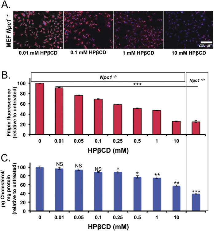
Figure 2. HPβCD removes cholesterol from Npc1−/− cells.
Npc1−/− MEFs were incubated with HPβCD (0–10 mM) for 24 hours. Cells were either fixed or lysed. (A) Fixed cells were imaged for filipin stain using high throughput imaging and representative images are show for 0.01, 0.1, 1 and 10 mM HPβCD treatments. (B) Filipin fluorescence per cell was quantified using Olympus scanR analysis software, and normalized to untreated cells. 48 images per treatment group were analyzed. (C) Cell lysates were analyzed for total cellular cholesterol using the amplex red assay and normalized by total protein content. Untreated Npc1+/+ MEFs are shown for comparison. Results were tested for statistical significance using one-way ANOVA with Tukey’s multiple comparison test. *P < 0.05, **P < 0.01, ***P < 0.001 and NS = not significant.

