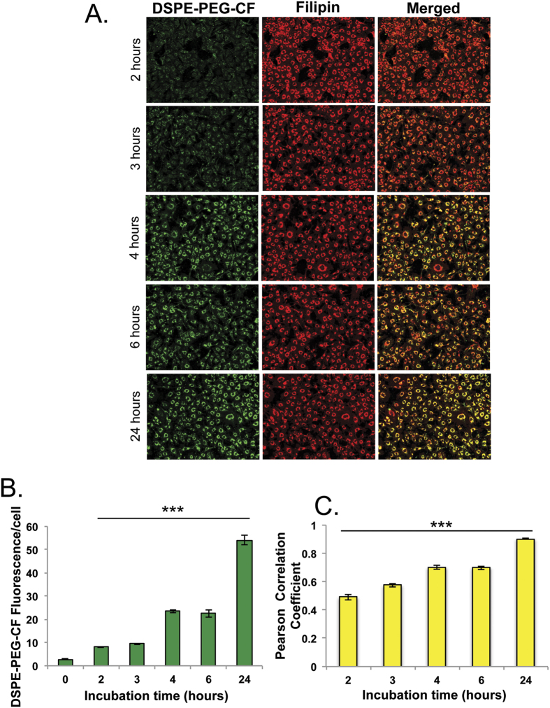
Figure 4. Labeled DSPE-PEG2k is internalized and localized to filipin positive vesicles in Npc1−/− cells.
Carboxy-Fluorescein (CF) labeled DSPE-PEG2k assembled into micelles by thin film rehydration was exposed to cells at 10 μM concentration for different time points (2, 3, 4, 6, 24 hours), cells were washed, fixed and filipin stained. (A) High throughput imaging (16 images/well) was performed to monitor DSPE-PEG2k-CF uptake and co-localization with endosomal cholesterol. (B) Image-J software was utilized to measure DSPE-PEG2k-CF uptake and (C) Monitor co-localization by calculation of Pearson Coefficient. (n = 5). Results were tested for statistical significance using one-way ANOVA with Tukey’s multiple comparison test relative to untreated cells (B) or 24 hour treated cells (C). *P < 0.05, **P < 0.01, ***P < 0.001 and NS = not significant.

