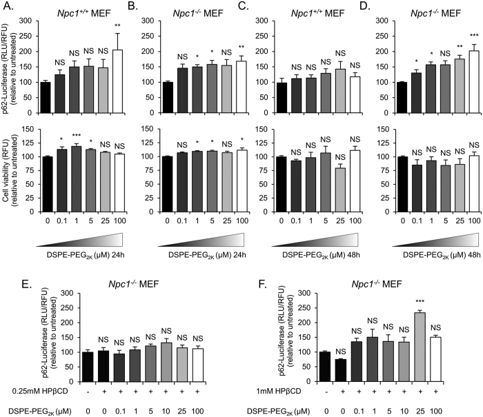
Figure 7. DSPE-PEG treatment slightly reduces autophagic degradation in Npc1+/+ and Npc−/− MEFs.
(A,C) p62-fluc.Npc1+/+ and, (B,D) p62-fluc. Npc−/− MEFs were induced to accumulate p62-fluc fusion protein with 1 μg/ml of Doxycycline for 24 hours. Doxycycline was washed from the cells and then they were treated with 0–100 μM DSPE-PEG2k for 24 (A and B upper panels) or 48 (C and D, upper panel) hours and autophagic degradation was measured using luciferase intensity. The cells were also analyzed for cell viability (lower panels). (E,F) Npc1−/− p62-fluc were also treated with a combination of HPβCD (0.25 mM or 1 mM, respectively) and DSPE-PEG (0–100 μM) for 24 hours after being induced for 24 hours with 1 μg/ml of Doxycycline. Results were tested for statistical significance using one-way ANOVA with Tukey’s multiple comparison test. *P < 0.05, **P < 0.01, ***P < 0.001 and NS = not significant.

