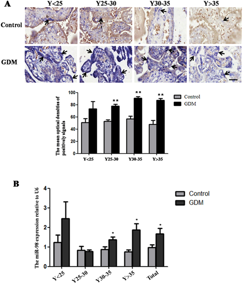
Figure 1. Up-regulation of miR-98 in the placental tissues from patients with GDM.
The expression of miR-98 in the placental tissues from patients with GDM and normal pregnant women was detected by in situ hybridization using DIG-labeled LNA probes specific to miR-98 (A). The stain was developed with BCIP/NBT. Black arrows indicate hybridization signals and the positive signals of miR-98 are blue. The scale bar indicates a distance of 50 μm. The histogram represents the MODs of positive signals of miR-98 in placentas. The expression of miR-98 in the placental tissues was also detected by qRT-PCR (B). U6 serves as an internal reference to normalize the experimental error. *P < 0.05; **P < 0.01.

